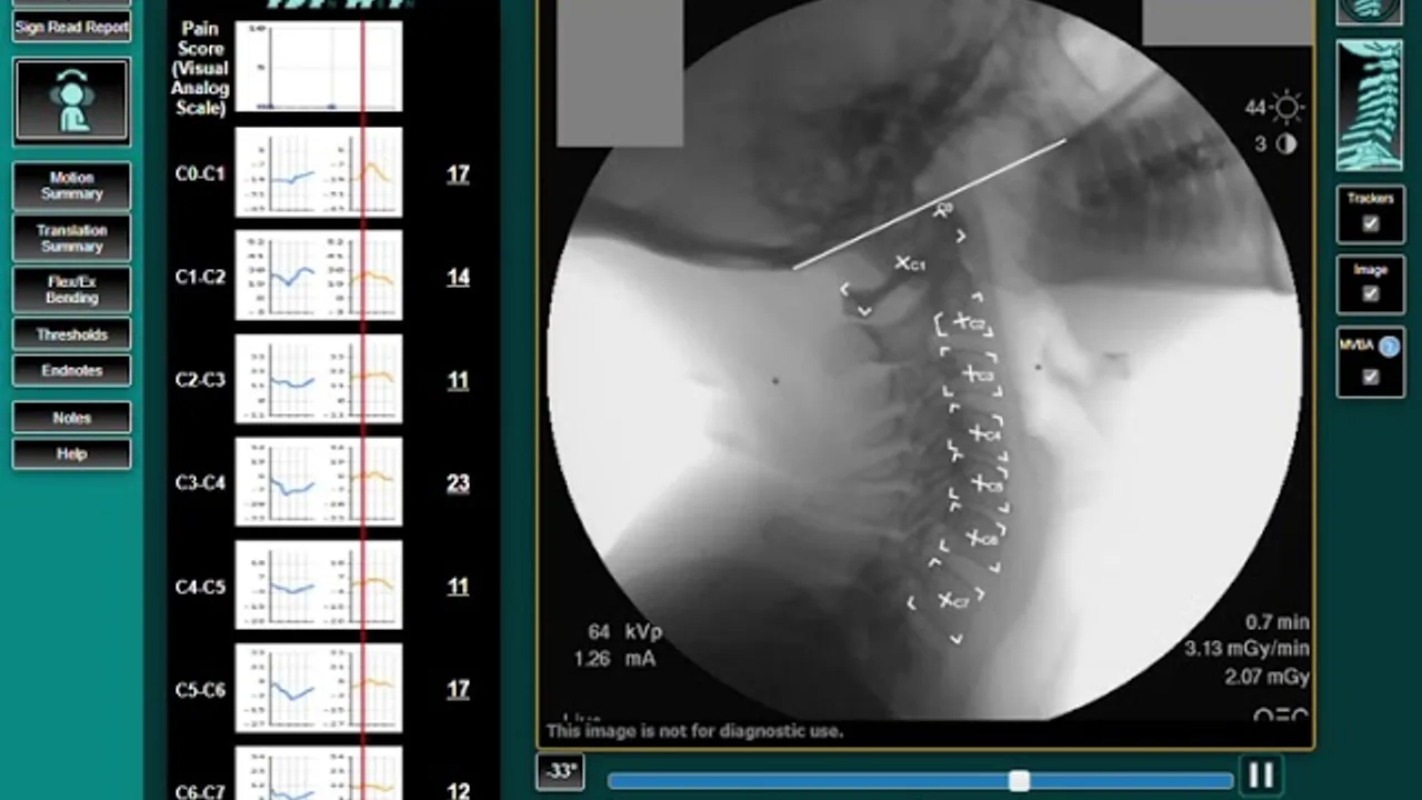
A still picture cannot show an unstable spine. Motion imaging measures what your spine does when it moves.
Why "unremarkable" imaging isn't the end of it
Your spine was holding perfectly still when it was photographed
That is the whole problem. A standard x-ray or MRI captures one motionless instant, and a torn or
stretched ligament is not a shape — it is a behaviour. Hold the joint still and it looks fine. Ask it
to move and it goes too far, or goes the wrong way. Flexion-extension films alone still miss that
instability in as many as 30% of cases.
Dr. Douglas DeSalvo is Imaging Facility Director of Precision Spine & Motion Imaging, in Suite 104 of our
own building — a facility built specifically to image the spine while it is working. He holds a
California x-ray operating permit, is a State of California Certified Fluoroscopy Owner-Operator, and
has post-graduate training in both radiology and MRI spine interpretation.
Vertebral Motion Analysis records the spine through controlled flexion and extension using pulsed fluoroscopy — in effect a movie of the spine in motion, rather than a photograph of it standing still.
Numbers, not impressions
Every vertebral level is measured: alignment, angulation, translation and disc height. The result is quantitative data an insurer, an attorney or a surgeon can evaluate on its own terms.
Catches what static films miss
Flexion-extension x-rays alone carry a false-negative rate of up to 30%. Motion analysis is designed for exactly the ligament instability those films let through.
Less radiation, wider eligibility
The FDA-cleared system uses up to 25% less radiation than a traditional four-view x-ray series, and is safe for patients with a pacemaker — who cannot have an MRI at all.
Independent reads
Studies are interpreted by a medical neuroradiologist, a third party, against scientific measurement criteria, so the report does not come from whoever is treating you.
Same building, one trip
Chiropractic care in Suite 108, imaging in Suite 104. A diagnosis does not cost you a second appointment across the county.
Who it's for
Three reasons a spine gets imaged in motion
Patients
If your pain does not match your imaging — or an MRI came back "unremarkable" while you still cannot turn your head — motion analysis often explains the gap.
Personal injury cases
Ligament injury is the crux of most collision claims and the hardest thing to demonstrate from static films. Motion analysis measures it.
Referring physicians & surgeons
Objective level-by-level instability data to support conservative management, or to select and plan a minimally invasive procedure.
Not every case needs a motion study. Most start with an examination and, when it will change the plan,
on-site digital x-ray — read the same day, in plain English. Motion analysis is the next step when the
question is specifically whether a segment has become unstable.
Digital x-ray on site at DeSalvo Chiropractic, Suite 108 — ordered only when clinically indicated
Vertebral Motion Analysis at Precision Spine & Motion Imaging, Suite 104
MRI review and interpretation, including studies taken elsewhere — bring the disc or the report
It is a motion-based imaging study. Instead of a single still exposure, patented equipment guides your neck or low back through a controlled range of movement while pulsed fluoroscopy records it at 8 frames per second. Software then measures how each vertebra moves relative to its neighbours, producing a level-by-level report of alignment, angulation, translation and disc height.
How is this different from the MRI I already had?+
An MRI is excellent at showing soft tissue at rest — discs, cord, nerve roots. What it cannot show is behaviour: whether a segment moves too far or in the wrong direction when you actually use it. Ligament injuries are functional problems, so they frequently look normal on a study taken while you hold still. The two tests answer different questions and are often used together.
Is it safe? How much radiation is involved?+
The system is FDA-cleared and uses pulsed rather than continuous fluoroscopy, which keeps the total dose up to 25% below a conventional four-view x-ray series. Because there is no magnetic field, it is also available to patients with a pacemaker or other implants that rule out MRI.
Does it hurt?+
No. The equipment moves you slowly and only as far as you are comfortable going, and you are in control the whole time. Most studies take a few minutes.
Do I need a referral?+
Talk to us first. Dr. Douglas DeSalvo will tell you whether motion imaging would actually change your care — and if it would not, he will say so. If it is indicated, the study is scheduled next door at Precision Spine & Motion Imaging.
Is it covered by insurance?+
Coverage varies by plan and by case, and on injury claims it is frequently billed through the claim rather than to you. Precision Spine & Motion Imaging handles its own billing — call them at (415) 432-9285 for specifics, or ask us and we will point you in the right direction.
Is Precision Spine & Motion Imaging part of DeSalvo Chiropractic?+
No — it is a separate diagnostic imaging facility in Suite 104 of the same building, and Dr. Douglas DeSalvo serves as its Imaging Facility Director. You can book imaging there without being a patient here, and vice versa.
Not sure whether you need it?
Start with an examination here. If a motion study would change your care we will tell you, and it is
scheduled next door. If it would not, we will tell you that too.